Preoperative detailed eye examination and consultation is essential for accurate vision correction. For an accurate measurement of corneal topography, patients should stop wearing soft lens for at least 1 week and the hard lens for at least 2~3 weeks prior to the test. Entrance of any foreign debris during surgery may cause infection and inflammation, so the patient should thoroughly wash face and avoid any eye makeup. Do not drink too much alcohol nor overwork the day before the surgery. It is also recommended to have sound sleep the night before. It is better not to drive right after the surgery, so it’d be advisable to accompany a guardian if bringing a car.
1. Definition
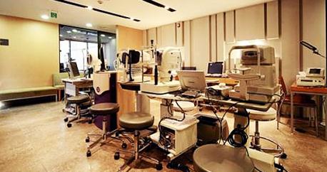
To correct your vision flawlessly without any complications, a thorough eye examination is required prior to the surgery.
2. Who are the ideal candidates for the examination?
- Those who wish to know if they can receive vision correction surgery.
- Those who have no plan to be pregnant within 6 months(You cannot get the surgery during pregnancy or breast feeding)
3. Thorough eye exam prior to the surgery

(*Some clinics may not perform DNA Test depending on the cases.)
Thorough Preoperative Eye Examinations
Auto Refracto-Keratometry
It measures the degrees of myopia, hyperopia and astigmatism, and automatically examines curvature radius and refraction of cornea.
Manifest Refraction with Skiascope
A patch of light is formed on the patient’s retina and by moving that patch in a given direction and observing the direction in which it appears to move after refraction by the patient’s eye, the retinoscopist can determine whether the patient’s retina is focused in front of, at, or behind the retinoscope’s sight hole.
Lensometry
It measures vergence and prism power of lens that a patient wears, and checks how far a patient’s eye sights can be corrected.
Best-Corrected Visual Acuity Test
It measures the best-corrected visual acuity of a patient. The presence of amblyopia can be examined through this test, and the degrees of post-treatment visual acuity can be predicted as well.
Test of Near Vision Accommodation Power
It analyzes the post-surgery near vision by testing the near vision and the accommodation power of an eye after correcting refraction abnormalities of myopia, hyperopia and astigmatism.
Cycloplegic Refraction
It is a procedure used to determine a person's refractive error by temporarily paralyzing the muscles that aid in focusing the eye. Cycloplegic eye drops are used to temporarily paralyze or relax the cililary body, or focusing muscle, of the eyes.
Wavefront Analysis
Wavefront-guided ablation is performed to measure irregularities in corneal surface for all patients who are subjected for the surgery.
ORBscan
It is a combination of reflective corneal topography and optical slit design that allows the analysis of the corneal thickness and posterior surface of the entire cornea. Those who have abnormalities in those areas or have thin cornea may not be able to have the surgery.
Topolyzer Test
It measures and generates high resolution treatment data of the corneal surface as well as its eccentricity and CO-WAVE abalation using Allegro-Topo - a corneal topographer to guide T-CAT LASIK surgery.
Q-Value Measurement
Custom-Q Treatment in surface ablation for myopic astigmatism, which provides the best postoperative visual acuity, is performed based on Q-value of a patient’s retina measured prior to the operation.
Ultrasound A-Scan
It provides data on the length of the eye and the power of the cornea (Keratometry), which is essential to calculate altisan lens power for altisan intraocular lens implant.
Pupil Diameter measurement
Pupil Diameter is measured in a dark room as a preoperative evaluation for refractive surgery to determine if the surgery is suitable or a wavefront operation is needed for a patient.
Schirmer’s Tear Test
It uses paper strips inserted into the eye for several minutes to measure the production of tears. It determines whether the eye produces enough tears to keep it moist.
Tear Film Break-Up Test
It is one of the dry eye tests which measures the time it takes for tears to break up in the eye after placing a drop of fluorescein in the cul-de-sac (under the corner of the lower eyelid).
Pachymetry
It measures the thickness of the cornea using ultrasound by comparing the value measured with ORBscan.
Tonometry
It determines the intraocular pressure (IOP), the fluid pressure inside the eye. It is an important test in the evaluation of patients with glaucoma. Normal IOP ranges from 10-21mmHg.
Visual Field Test
It analyzes a patient’s visual field using Humphrey field exam, an industrial standard tester. This test distinguishes glaucoma or retinal disorder.
Dominant Eye Test
It determines which eyes is dominant, which is an important consideration for monovision correction to reduce the need for reading glasses or bifocals.
Prism Cover-Uncover Test
It ascertains if the patient has eso- or exo- tropia or phoria and its.
Slit Lamp Examination
It examines the anterior segment, or frontal structures and posterior segment of the human eye, which includes the eyelid, sclera, conjunctiva, iris, natural crystalline lens, and cornea.
Funduscopy
It examines the internal part of an eye such as optic disc, retina and blood vessels, which is essential to diagnose conditions that cause increased intracranial pressure like glaucoma, diabetes, hypertension.
Stereopsis Test
It examines the visual perception of depth from the two slightly different projections of the world onto the retinas of the two eyes.
Contrast Sensitivity Test
It determines how well a patient’s eyes function in low light and how well he/she can distinguish objects from similarly colored or shaded backgrounds.
Avellino DNA Test
It detects Avellino Corneal Dystrophy. Avellino Corneal Dystrophy is a rare inherited eye disorder in which abnormal material often accumulates in the clear (transparent) outer layer of the eye (cornea). It is often progressive and affects both eyes (bilateral) which may lead to degeneration of the cornea.
KMH Recommended Tips
Exercise Tips for Good Vision
No.8 Eye Exercise
- At first, make a number 8 shape with your thumbs and index fingers.
- Follow the shape by rolling your eyes as large as possible.
Butterfly Eye Exercise
- Roll your eyes in butterfly shape using the method described above.
Up&Down Eye Exerscise
- Roll your eyes to the direction of 12 & 6 o’clock in fixed position.
Left & Right Eye Exerscise
- Roll your eyes to the direction of 9 & 3 o’clock.
- After the Left&Right exercise, try diagonal exercise to the direction of 11, 7, 1 & 7 o’clock.
Circular Excercise
- Roll your eyes clockwise and counter-clockwise repeatedly from the direction of 6 o’clock.
- Then roll your eyes to draw a big circle from the inside toward the outside of the body for visual accommodation and rotational exercises.